Rylea Taylor was in a head-on collision car accident with her two children in the car. While miraculously she and her daughter were okay, when she pulled her 16-month-old son Jaxon out of the wrecked car, she knew immediately it was bad. Jaxon’s neck was broken. Her infant son had suffered what is called internal decapitation, from which normally the best-case scenario is paralysis. Incredibly, a few months later, Jaxon was walking as if nothing had happened. This is how doctors reattached the little boy’s head.
Doctor’s Successfully Reattached Baby’s Head After Internal Decapitation
Internal decapitation sounds gruesome, and rightfully so. Most who suffer this, often seen in car-crash scenarios, don’t make it out alive. Little baby Jaxon’s case, however, can only be described as a combination of a talented surgeon, modern medicine, and a lot of luck. Jaxon was in a head-on collision going 70 miles per hour at less than two years of age. While his car seat kept his body strapped in securely, his head was whipped in such a way that he fractured his top two vertebrae and tore apart the ligaments that stabilized them. This meant that his top two vertebrae and skull were completely detached from his neck and spinal column. (1)
As already mentioned, this usually means near-immediate death, if not complete paralyzation. This is because the pathway between the brain and lungs is usually completely severed, meaning the victim cannot breathe unaided. Miraculously, however, Jaxon was still breathing on his own. Thankfully, Jaxon landed in the care of a senior spinal surgeon at Brisbane, Australia’s Lady Cilento Children’s Hospital Geoffrey Askin, also known as Australia’s Godfather of Spinal Surgery.
A Dire Situation
As already stated, most people don’t survive an internal decapitation, medically known as a C1-C2 dislocation. One study found that 68% of people perish at the site of the accident before their dislocation is even diagnosed, and another 22% die in the hospital. Of those who do survive, they are left completely paralyzed and usually unable to breathe unassisted. They are most common in young children because their heads are much heavier than their necks are capable of stabilizing in those situations. (2)
Diagnosing the dislocation is also complicated. You have to avoid moving the patient as much as possible, but most traditional imaging techniques require you to do so. In Jaxon’s case, doctors at a hospital near the crash site first put Jaxon inside a CT scanner. This allowed the x-ray beam to rotate around him, giving the doctors a 3D image of his injury. The staff at the first hospital sent those to Askin at Lady Cilento while Jaxon was airlifted there. While the injury was extreme, somehow, the nerves in the spinal cord had remained intact, which is why Jaxon could continue to breathe unassisted.
The doctors at Lady Cilento then gave Jaxon an MRI to further examine the extent of his injury. This allowed them to quickly construct and prepare all necessary tools and braces that would be required for the surgery and Jaxon’s recovery. The next day, Askin and his team of 20 doctors were ready for the six-hour surgery that lay ahead.
The Surgery
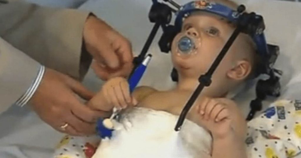
The doctors had to create a custom-made “halo” brace for Jaxon, both to hold his head in place during the surgery and for the three-month post-op recovery period. The surgery’s first step is to realign Jaxon’s skull and neck. Once the broken vertebrae and spinal cord are in the correct position, they reattach the two.
While many advancements have been made in this area, there are always things you don’t know until the patient is on the operating table. Because most patients don’t even make it out of the crash site, let alone to the hospital, these surgeries are rarely done. Normally the reattachment is done using surgical screws. However, Jaxon’s vertebrae were too small for even the smallest version of these. Instead, they had to use the very old technique of using a wire and then graft a fragment from one of Jaxon’s ribs onto the joint to provide strength that the wire does not have.
A Success
Though long and grueling, the surgery was successful. Just three weeks later, Jaxon could return to his hometown and was even toddling alone as a toddler should, using just one of his parents’ fingers as a bit of extra support. He continued to wear the halo brace for three months and made regular trips to his local hospital for x-rays. Askin used these x-rays to be sure that everything was staying in the position that it should and that he was recovering well.
Askin says that Jaxon is incredibly lucky, as most who suffer those injuries don’t have the same outcome. For the most part, he will be able to grow up living a normal life. He will only have to avoid activities like playing Rugby, which could cause whiplash and damage the injury site.
Keep Reading: 3-Year-Old Conjoined Twins With Fused Brains Separated in Historic Virtual Reality Surgery
Sources
- How Surgeons Reattached a Toddler’s Head.” Scientific America. K. J. Lee. October 19, 2015.
- “Identifying survivors with traumatic craniocervical dissociation: a retrospective study.” PubMed. Zara Cooper, Joel A Gross, J Matthew Lacey, Neal Traven, Sohail K Mirza, Saman Arbabi. May 2009.